

Abstract
Objective: Juvenile idiopathic inflammatory myopathies (JIIM) exhibit heterogeneous presentation, and intravenous immunoglobulin (IVIG) is often required in refractory cases. This study aimed to identify the clinical characteristics associated with IVIG use.
Methods: This retrospective study included patients aged 0–18 years who fulfilled the Bohan and Peter and/or 2017 EULAR/ACR criteria for JIIM and were followed between March 2009 and August 2025. Demographic, clinical, laboratory, and treatment data were recorded.
Results: Thirty-eight patients were included; 29 (76.3%) had juvenile dermatomyositis. Sixteen patients received IVIG. Severe or unresponsive muscle weakness was the main indication (56.3%). IVIG-treated patients had significantly lower Childhood Myositis Assessment Scale (CMAS) scores at diagnosis and at the third month, higher AST levels, more frequent nailfold capillaroscopy abnormalities, and lower remission rates. Each one-point decrease in CMAS increased the odds of IVIG use. Additionally, the third-month CMAS score was the only independent predictor for IVIG therapy.
Conclusion: In conclusion, IVIG is an effective treatment option for patients with more severe and refractory JIIM. These patients are characterized by lower CMAS scores and marked microvascular involvement. Earlier use of IVIG as a steroid-sparing strategy may help restrict adverse effects.
Keywords: juvenile dermatomyositis, myositis, intravenous immunoglobulins, capillaroscopy
INTRODUCTION
Juvenile idiopathic inflammatory myopathies (JIIM) are rare pediatric autoimmune conditions characterized by immune-mediated myositis and a prominent microvascular vasculopathy.1 The annual incidence of JIIM is estimated to be 2-4 per million children.2 Various subtypes have been identified within JIIM, including juvenile dermatomyositis (JDM), juvenile polymyositis, inclusion body myositis, and amyopathic dermatomyositis.3 JDM, which accounts for approximately 80–85% of all JIIM cases.4
The first-line treatment of JIIM usually consists of immunosuppressants such as glucocorticoids and methotrexate. Some patients may not achieve disease control with those and may require additional treatments.5 IVIG is often used in combination with disease-modifying antirheumatic drugs (DMARDs) to induce and maintain remission.6,7 Current studies have demonstrated that IVIG is an effective and safe treatment option for patients with severe or treatment-refractory JDM.8,9
The pathogenesis of JIIM is complex and multifactorial, driven predominantly by type I interferon activation, capillary dropout, complement deposition, and endothelial activation, which together contribute to a characteristic vasculopathy.5 The therapeutic effects of IVIG are considered to result from the neutralization of autoantibodies, the attenuation of microangiopathy, and the suppression of the type I interferon-mediated inflammatory response in JDM.10
The present study aims to describe the clinical, laboratory, and treatment characteristics of children with JIIM who received IVIG therapy and to identify the clinical factors that warranted IVIG treatment.
MATERIALS AND METHODS
Patient selection
The study included the data on patients aged 0–18 years who met the Bohan and Peter classification criteria and/or the 2017 European Alliance of Associations for Rheumatology/American College of Rheumatology (EULAR/ACR) classification criteria for JIIM retrospectively and who were followed up in the pediatric rheumatology department between March 2009 and August 2025.11-13 Patients with missing data and a follow-up duration less than 12 months were excluded.
Data collection
Demographic characteristics, clinical manifestations, laboratory results, treatment, and disease outcomes were collected from patient records. Patient data for IVIG therapy were recorded in detail.
Definitions
Disease activity and treatment response were evaluated through clinical examination and serial measurements of serum muscle enzyme levels. The Childhood Myositis Assessment Scale (CMAS) was used to assess muscle strength during follow-up.14
Clinically inactive disease was defined according to the PRINTO criteria, requiring at least three of the following: creatine kinase ≤150 U/L, CMAS ≥48, Manual Muscle Testing of eight muscle groups (MMT8) ≥78, and physician’s global assessment ≤0.2 cm on a 10-cm visual analog scale.15
Refractory JIIM was defined as patients with an inadequate clinical response to first-line treatments such as corticosteroids and methotrexate or another immunosuppressive agent.16
Treatment protocol
In our clinic, IVIG therapy was administered to patients with JIIM who had ongoing active disease despite standard treatment with corticosteroids and/or conventional DMARDs. The decision to initiate IVIG therapy was guided by the clinician’s overall evaluation of disease activity and accompanying clinical findings. In these patients, IVIG was administered in combination with conventional and/or biologic DMARDs at a total monthly dose of 2 g/kg, with a maximum single dose of 70 g.
Ethical approval
The study was approved by the local ethics committee (approval no: TABED 2-25-1672). It was conducted in accordance with the ethical standards of the institutional and national research committees and the principles of the Declaration of Helsinki.
Statistical analysis
The research data were evaluated using the Statistical Package for the Social Sciences (SPSS) version 23, IBM Corp., Armonk, NY, USA, a statistical package program. In the descriptive statistics section, categorical variables were presented in tables by giving numbers and percentages, and continuous variables were presented with the median (IQR). The normality of continuous variables was evaluated using visual methods (histograms and probability plots) and analytical methods (Kolmogorov–Smirnov/Shapiro–Wilk tests). The Mann–Whitney U test was used to compare continuous variables that did not conform to a normal distribution between two groups. The chi-square test was used to compare categorical variables. The statistical significance level was accepted as p<0.05. Potential collinearity between explanatory variables was assessed prior to regression analyses. Variance inflation factors (VIFs) and correlation matrices were examined to identify overlapping effects. When variables exhibited high collinearity (VIF > 5 or strong pairwise correlations), only the clinically more relevant variable was retained in the multivariate model to avoid instability of the estimates. Variables with a p-value less than 0.25 in the bivariate comparisons were included in the bivariate regression analysis. Variables that were significant in the bivariate regression analysis were then entered into the multivariate logistic regression model. We used the “backward” selection method to identify the most significant variables in the final model.
RESULTS
A total of 38 patients diagnosed with JIIM were included in the study, of whom 23 (60.5%) were female. The median age at symptom onset was 8.5 years (IQR: 6.8–11.3), and the median age at diagnosis was 9 years (IQR: 6.0–12.3), respectively. The median interval between symptom onset and diagnosis was 2 months (IQR: 1.0–6.5), and the median follow-up period was 31 months (IQR: 13.3–59).
Of the 38 patients, 29 (76.3%) had JDM. Five patients (13.2%) had the amyopathic type, three patients (7.9%) had the hypomyopathic type, and one patient (2.6%) had immune-mediated necrotizing myopathy.
Muscle weakness was present in 31 patients (81.6%), and cutaneous manifestations in 35 (92.1%). Among the cutaneous findings, Gottron’s papules were observed in 25 patients (65.8%), heliotrope rash in 27 (71.1%), V-sign in 10 (26.3%), shawl sign in 8 (21.1%), and ulcerative lesions in 6 (15.8%) patients. Extramuscular manifestations were arthritis (13, 34.2%), gastrointestinal involvement (n=9, 23.7%), dysphagia (n=7, 18.4%), dysphonia (n=5, 13.2%), cardiac involvement (n=1, 2.6%), and pulmonary involvement (n=1, 2.6%) (Table 1).
| bMedian (IQR), IQR: interquartile range, αChi-square test, ¥Mann-Whitney U test, β n (%) | ||||
| Table 1. The comparison of the demographic and clinical characteristics of JIIM patients according to IVIG therapy | ||||
|
|
|
|
|
|
| Age, yearsb |
|
|
|
|
| Sex, femaleβ |
|
|
|
|
| Age at symptom onset, yearsb |
|
|
|
|
| Age at diagnosis, yearsb |
|
|
|
|
| Time from symptom onset to diagnosis, monthsb |
|
|
|
|
| Follow-up duration, monthsb |
|
|
|
|
| Clinical findingsβ | ||||
| Muscle weakness |
|
|
|
|
| Constitutional symptoms |
|
|
|
|
| Cutaneous involvement | ||||
| Rash |
|
|
|
|
| Gottron’s papule |
|
|
|
|
| Heliotrope rash |
|
|
|
|
| V-sign |
|
|
|
|
| Shawl sign |
|
|
|
|
| Arthritisβ |
|
|
|
|
| Cardiac involvement |
|
|
|
|
| Pulmonary involvement |
|
|
|
|
| Gastrointestinal involvement |
|
|
|
|
| Dysphagia |
|
|
|
|
| Dysphonia |
|
|
|
|
| Subcutaneous edema |
|
|
|
|
| Lipodystrophy |
|
|
|
|
The median CMAS score at the initial presentation was 32.5 (IQR: 21.0–45.3). By the 3-month follow-up, this had increased to 43.0 (IQR: 38.3–46.5). It further improved to 46.0 (IQR: 42.5–50.0) at 6 months and to 51.0 (IQR: 50.0–52.0) by the final follow-up (Table 2).
| bMedian (IQR); IQR: interquartile range; αChi-square test; ¥Mann-Whitney U test; β n (%); CMAS: Childhood Myositis Assessment Scale; AST: Aspartate Aminotransferase; LDH: Lactate Dehydrogenase; CK: Creatine Kinase; JIIM: Juvenile Idiopathic Inflammatory Myopathies; MSA: Myositis-Specific Antibodies; Anti-NXP2; Anti–nuclear matrix protein 2; Anti-SRP: Anti–signal recognition particle; Anti-TIF1 (TIF1-γ): Anti–transcription intermediary factor 1 gamma; Anti-Mi-2: Anti–Mi-2 nucleosome remodeling deacetylase complex; Anti-MDA5: Anti–melanoma differentiation–associated gene 5; Anti-SAE: Anti–small ubiquitin-like modifier activating enzyme; Anti-PL-7: Anti–threonyl–tRNA synthetase; Anti-OJ: Anti–isoleucyl–tRNA synthetase | ||||
| Table 2. The comparison of the laboratory, treatment, and outcome characteristics of JIIM patients according to IVIG therapy | ||||
|
|
|
|
|
|
| AST, U/Lb |
|
|
|
|
| LDH, U/Lb |
|
|
|
|
| CK, U/Lb |
|
|
|
|
| MSA positivity |
|
|
|
|
| Anti-NXP2 |
|
|
|
|
| Anti-SRP |
|
|
|
|
| Anti-TIF1-γ |
|
|
|
|
| Anti -Mi-2 |
|
|
|
|
| Anti-MDA5 |
|
|
|
|
| Anti-SAE |
|
|
|
|
| Anti-PL-7 |
|
|
|
|
| Anti-OJ |
|
|
|
|
| Disease Activityb | ||||
| CMAS at presentation |
|
|
|
|
| CMAS at 3 months |
|
|
|
|
| CMAS at 6 months |
|
|
|
|
| CMAS at last visit |
|
|
|
|
| Nailfold capillary abnormalitiesβ |
|
|
|
|
| Treatmentβ | ||||
| Pulse methylprednisolone |
|
|
|
|
| Methylprednisolone (2 mg/kg/day) |
|
|
|
|
| Methotrexate |
|
|
|
|
| Mycophenolate mofetil |
|
|
|
|
| Hydroxychloroquine |
|
|
|
|
| Cyclophosphamide |
|
|
|
|
| Prognosisβ | ||||
| Remission |
|
|
|
|
| Relapse |
|
|
|
|
| Refractory JIIM |
|
|
|
|
Myositis-specific autoantibody positivity (MSA) was present in 21 patients (55.3%); 8 patients (21.1%) were negative, and 9 patients (23.7%) were not tested. Among the 21 patients with MSA positivity, NXP2 was the most frequently detected antibody in 9 patients (31.0%). This was followed by the presence of TIF1 antibodies in 5 patients (17.2%) and MDA5 antibodies in 3 patients (10.3%). Antinuclear antibody (ANA) positivity was present in 18 patients (47.4%).
Characteristics of patients who received intravenous immunoglobulin treatment
Ten of the 16 patients (62.5%) received IVIG at the time of diagnosis. During the follow-up period, eight patients (50%) required IVIG therapy. Two patients (12.5%) received IVIG therapy in the first three months, and four patients (25%) received it in the second three months. Two patients required a second course of IVIG due to persistent calcinosis and muscle weakness.
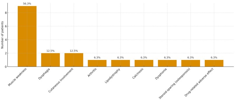
The reasons for IVIG administration were severe or unresponsive muscle weakness (56.3%), dysphagia (12.5%), persistent cutaneous involvement (12.5%), arthritis (6.3%), lipodystrophy (6.3%), calcinosis (6.3%), and dysphonia (6.3%). In addition, IVIG was initiated owing to a drug-related adverse effect (6.3%) and as a steroid-sparing strategy (6.3%) due to steroid-induced osteoporosis. The indications for IVIG therapy are shown in Figure 1.
Adverse effects of IVIG therapy were observed in two patients: hemolysis and severe headache.
Comparison of patients with/without intravenous immunoglobulin therapy
Comparison of demographic, laboratory, treatment, and outcome characteristics of JIIM patients according to IVIG treatment is presented in Tables 1 and 2. There were no significant differences between the two groups in demographic and clinical characteristics. AST levels were significantly higher in the IVIG-treated group. In terms of the presence of MSA, there was no significant difference between the two groups.
The CMAS score at onset was significantly lower in IVIG-treated patients (p=0.021). By the 3-month follow-up, CMAS remained lower in the IVIG-treated group (p=0.011). No significant differences were found at the sixth month (p=0.109) and the last visit (p=0.753).
Abnormalities in nailfold capillaroscopy (NFC) at onset were significantly more common in IVIG-treated patients (p = 0.039).
The remission rate was significantly lower in the IVIG-treated patients (p=0.005). All IVIG-treated patients had refractory JIIM, although this finding was not statistically significant.
Predictors of intravenous immunoglobulin therapy
In the bivariate logistic regression analysis, lower CMAS scores were significantly associated with an increased odds of receiving IVIG therapy. At diagnosis, each one-point decrease in the CMAS score was associated with a 1.09-fold increase in the odds of IVIG administration (OR: 0.92; 95% CI: 0.87–0.97; p=0.007). Similarly, at the third month, each one-point decrease corresponded to a 1.30-fold increase in IVIG use (OR: 0.77; 95% CI: 0.60–0.98; p=0.038). In the multivariate model, the third-month CMAS score remained the only variable significantly associated with IVIG therapy, with each one-point decrease increasing the odds of IVIG administration by approximately 1.30-fold (OR: 0.77; 95% CI: 0.60–0.97; p=0.038) (Table 3).
| Hosmer and Lemeshow p=0.568, OR: odds ratio, CI: confidence interval, CMAS: Childhood Myositis Assessment Scale | ||||
| Table 3. Multivariate logistic regression model for predicting IVIG therapy of JIIM | ||||
| Factor |
|
|
|
|
| CMAS score at diagnosis |
|
|
||
| CMAS score at 3rd month |
|
|
|
|
DISCUSSION
Juvenile idiopathic inflammatory myopathies are a group of diseases with marked clinical heterogeneity that complicates management. Therefore, an individualized treatment approach is required to reduce morbidity and achieve disease control. Our study demonstrated that patients receiving IVIG had lower CMAS scores and more frequent abnormalities in NFC. Furthermore, despite receiving pulse methylprednisolone and mycophenolate mofetil, the remission rate was lower in patients receiving IVIG.
According to the EULAR/ACR and SHARE recommendations, the therapeutic strategy for JIIM is guided by disease severity. For mild disease, the standard first-line therapy is methotrexate in combination with oral corticosteroids. For moderate-to-severe cases, or when the initial treatment is ineffective, high-dose intravenous corticosteroids are administered alongside methotrexate. IVIG is recommended as an adjunctive therapy for patients with refractory disease, frequent relapses, or prominent cutaneous involvement.17-19 In our study, disease activity, assessed using the CMAS at diagnosis and at the third-month visit, was higher in patients requiring IVIG treatment. Additionally, a higher AST level at onset was defined in the IVIG-treated group. Similarly, Lam et al. reported that patients receiving IVIG had more severe muscle involvement, lower MMT scores, and more frequent dysphagia and photosensitivity than the control group.8 In our study, logistic regression analysis demonstrated that lower CMAS scores were associated with IVIG use. Likewise, severe initial muscle weakness and persistent high disease activity despite first-line treatment were more frequently observed in patients who received IVIG. All these findings may suggest both more severe disease activity and clinicians’ tendency to prefer IVIG treatment based on this perceived severity.
Recent studies have shown that early aggressive immunosuppressive therapy, such as pulse methylprednisolone and/or IVIG therapy in the first year, provided better clinical response and faster corticosteroid tapering.20,21 A pediatric study reported that using IVIG in the early period reduced steroid burden, improved refractory muscle weakness and persistent cutaneous findings.22 Lam et al. found that patients receiving IVIG treatment who had more severe disease at baseline achieved remission slowly. IVIG was shown to significantly reduce disease activity, particularly in steroid-resistant patients.8 The CARRA study also demonstrated that most clinicians add IVIG early in the management of patients, particularly those who are refractory to treatment, steroid-dependent, or experiencing toxicity, and reported significant clinical improvement in severe phenotypes such as refractory muscle weakness, ulceration, and calcinosis.16,23 In the present study, we observed that all patients receiving IVIG therapy had a refractory JDM phenotype, and remission rates were lower in this group.
In JIIM, strict sun protection is an indispensable non-pharmacological component of treatment. It is a fundamental preventive measure in disease management. Furthermore, there is a long-term use of corticosteroids, which are the mainstay of treatment. The possibility of adverse effects increases because of the steroid burden.24 Hence, the use of steroid-sparing agents is important to reduce complications. It has previously been reported that IVIG therapy is also used as a steroid-sparing agent in JIIM.25,26 In our study, IVIG was added in one patient due to secondary osteoporosis as a steroid-sparing strategy, and in another due to drug-related adverse effects. IVIG may play an important role in individualized treatment approaches, not only for refractory cases but also for steroid sparing.
Nailfold capillaroscopy abnormalities play a crucial role in assessing early changes in vasculopathies. The NFC in JIIM reflects microvascular damage and disease activity. Visualizing early microvascular damage, capillary loss, and neoangiogenic patterns can provide clinicians with insight into disease severity. Schmeling et al. reported that pathogenic NFC is associated with both muscle and skin disease activity.27 Barth et al. demonstrated that a decreased capillary count and neoangiogenesis in NFC are associated with higher disease activity and pulmonary involvement.28 In some studies, a significant reduction in NFC density was found in children with JDM, but no association was observed with CMAS scores.29,30 In our study, NFC abnormalities were more frequently observed in the IVIG-treated group. Given these results, NFC appears to be a complementary tool in classifying early disease activity. Furthermore, early-phase NFC findings may indicate that the patients require IVIG.
The limitations of this study are its retrospective design and small sample size. As a tertiary care center, although our patients had high disease severity and serious organ involvement, all patients who met the inclusion criteria were included in the study to prevent bias. One limitation of the study is that MSA tests are not routinely performed at our center, so they could not be evaluated in all patients; this may have limited the analysis of the relationship between autoantibody profiles and clinical findings. IVIG initiation was based on clinician assessment rather than standardized criteria, and IVIG preparations varied according to hospital availability.
In conclusion, IVIG is an effective treatment option for patients with severe and refractory JIIM. These patients are characterized by lower CMAS scores and marked microvascular involvement. Therefore, NFC can be considered an important.
Ethical approval
This study was approved by the Bilkent City Hospital Medical Research Ethics Committee (TABED) (Date: 26.11.2025, Decision/Protocol No: TABED 2-25-1672). Informed consent was obtained from all participants involved in this study.
Data availability statement
The data that support the findings of this study are available from the corresponding author upon reasonable request.
Conflict of interest
The authors declare that this study was conducted in the absence of any commercial or financial relationships that could be construed as a potential conflict of interest.
Funding
The authors declare that this study received no funding.
Generative AI statement
The authors declare that no generative AI or AI-assisted technologies were used in the writing or preparation of this study.
References
- Pilkington C, Feldman B, Sontichai W. Juvenile dermatomyositis and other inflammatory muscle diseases. In: Petty RE, Laxer RM, Lindsley CB, Wedderburn LR, Mellins ED, Fuhlbrigge RC, editors. Textbook of Pediatric Rheumatology. 8th ed. Philadelphia: Elsevier; 2021: 360-376.
- Meyer A, Meyer N, Schaeffer M, Gottenberg JE, Geny B, Sibilia J. Incidence and prevalence of inflammatory myopathies: a systematic review. Rheumatology (Oxford). 2015;54:50-63. https://doi.org/10.1093/rheumatology/keu289
- Rider LG, Nistala K. The juvenile idiopathic inflammatory myopathies: pathogenesis, clinical and autoantibody phenotypes, and outcomes. J Intern Med. 2016;280:24-38. https://doi.org/10.1111/joim.12444
- Cancarini P, Nozawa T, Whitney K, et al. The clinical features of juvenile dermatomyositis: a single-centre inception cohort. Semin Arthritis Rheum. 2022;57:152104. https://doi.org/10.1016/j.semarthrit.2022.152104
- Papadopoulou C, Chew C, Wilkinson MGL, McCann L, Wedderburn LR. Juvenile idiopathic inflammatory myositis: an update on pathophysiology and clinical care. Nat Rev Rheumatol. 2023;19:343-62. https://doi.org/10.1038/s41584-023-00967-9
- Marrani E, Abu-Rumeileh S, Mastrolia MV, Maccora I, Pagnini I, Simonini G. A systematic review on biological therapies in juvenile idiopathic inflammatory myopathies: an evidence gap in precision medicine. Clin Exp Rheumatol. 2022;40:457-70. https://doi.org/10.55563/clinexprheumatol/ltrj4l
- Sener S, Cam V, Ozen S, Batu ED. Biologic drugs in the treatment of juvenile dermatomyositis: a literature review. Clin Rheumatol. 2024;43:591-602. https://doi.org/10.1007/s10067-023-06740-3
- Lam CG, Manlhiot C, Pullenayegum EM, Feldman BM. Efficacy of intravenous Ig therapy in juvenile dermatomyositis. Ann Rheum Dis. 2011;70:2089-94. https://doi.org/10.1136/ard.2011.153718
- Doudouliaki T, Papadopoulou C, Deakin CT. Use of rescue therapy with IVIG or cyclophosphamide in juvenile myositis. Curr Rheumatol Rep. 2021;23:24. https://doi.org/10.1007/s11926-021-00990-3
- Gandiga PC, Ghetie D, Anderson E, Aggrawal R. Intravenous immunoglobulin in idiopathic inflammatory myopathies: a practical guide for clinical use. Curr Rheumatol Rep. 2023;25:152-168. https://doi.org/10.1007/s11926-023-01105-w
- Bohan A, Peter JB. Polymyositis and dermatomyositis (first of two parts). N Engl J Med. 1975;292:344-7. https://doi.org/10.1056/NEJM197502132920706
- Bohan A, Peter JB. Polymyositis and dermatomyositis (second of two parts). N Engl J Med. 1975;292:403-7. https://doi.org/10.1056/NEJM197502202920807
- Lundberg IE, Tjärnlund A, Bottai M, et al. 2017 European League Against Rheumatism/American College of Rheumatology classification criteria for adult and juvenile idiopathic inflammatory myopathies and their major subgroups. Arthritis Rheumatol. 2017;69:2271-2282. https://doi.org/10.1002/art.40320
- Huber AM, Feldman BM, Rennebohm RM, et al. Validation and clinical significance of the Childhood Myositis Assessment Scale for assessment of muscle function in the juvenile idiopathic inflammatory myopathies. Arthritis Rheum. 2004;50:1595-603. https://doi.org/10.1002/art.20179
- Lazarevic D, Pistorio A, Palmisani E, et al. The PRINTO criteria for clinically inactive disease in juvenile dermatomyositis. Ann Rheum Dis. 2013;72:686-93. https://doi.org/10.1136/annrheumdis-2012-201483
- Spencer CH, Rouster-Stevens K, Gewanter H, et al. Biologic therapies for refractory juvenile dermatomyositis: five years of experience of the childhood arthritis and rheumatology research alliance in North America. Pediatr Rheumatol Online J. 2017;15:50. https://doi.org/10.1186/s12969-017-0174-0
- Liu K, Tomlinson G, Reed AM, et al. Pilot study of the juvenile dermatomyositis consensus treatment plans: a CARRA registry study. J Rheumatol. 2021;48:114-122. https://doi.org/10.3899/jrheum.190494
- Bellutti Enders F, Bader-Meunier B, Baildam E, et al. Consensus-based recommendations for the management of juvenile dermatomyositis. Ann Rheum Dis. 2017;76:329-40. https://doi.org/10.1136/annrheumdis-2016-209247
- Kobayashi I. Advances in juvenile dermatomyositis: pathophysiology, diagnosis, treatment and interstitial lung diseases-a narrative review. Children (Basel). 2024;11:1046. https://doi.org/10.3390/children11091046
- Hoff LS, de Souza FHC, Miossi R, Shinjo SK. Long-term effects of early pulse methylprednisolone and intravenous immunoglobulin in patients with dermatomyositis and polymyositis. Rheumatology (Oxford). 2022;61:1579-88. https://doi.org/10.1093/rheumatology/keab597
- Barut K, Aydin POA, Adrovic A, Sahin S, Kasapcopur O. Juvenile dermatomyositis: a tertiary center experience. Clin Rheumatol. 2017;36:361-366. https://doi.org/10.1007/s10067-016-3530-4
- Orandi AB, Fotis L, Lai J, et al. Favorable outcomes with reduced steroid use in juvenile dermatomyositis. Pediatr Rheumatol Online J. 2021;19:127. https://doi.org/10.1186/s12969-021-00615-0
- Reyna-Juárez Y, Mejía-Domínguez NR, Ostos-Prado MJ, et al. The Refractory DermatoMyositis Index (ReDMI): a clinical tool to predict refractory disease in patients with dermatomyositis. Clin Exp Rheumatol. 2026;44:289-96. https://doi.org/10.55563/clinexprheumatol/k77xaj
- Marstein HS, Godang K, Flatø B, Sjaastad I, Bollerslev J, Sanner H. Bone mineral density and explanatory factors in children and adults with juvenile dermatomyositis at long term follow-up; a cross sectional study. Pediatr Rheumatol Online J. 2021;19:56. https://doi.org/10.1186/s12969-021-00543-z
- Campanilho-Marques R, Fonseca JE, Machado PM. Treatment of idiopathic inflammatory myopathies. Joint Bone Spine. 2025;92:105932. https://doi.org/10.1016/j.jbspin.2025.105932
- Yeşilbaş O, Yıldız M, Yozgat CY, et al. A fatal interstitial lung disease in an anti-melanoma differentiation-associated gene 5 (anti-MDA5) antibody negative patient with juvenile dermatomyositis. Turk J Pediatr. 2021;63:903-8. https://doi.org/10.24953/turkjped.2021.05.018
- Schmeling H, Stephens S, Goia C, et al. Nailfold capillary density is importantly associated over time with muscle and skin disease activity in juvenile dermatomyositis. Rheumatology (Oxford). 2011;50:885-93. https://doi.org/10.1093/rheumatology/keq407
- Barth Z, Witczak BN, Flatø B, Koller A, Sjaastad I, Sanner H. Assessment of microvascular abnormalities by nailfold capillaroscopy in juvenile dermatomyositis after medium- to long-term followup. Arthritis Care Res (Hoboken). 2018;70:768-776. https://doi.org/10.1002/acr.23338
- Doğantan Ş, Taşkın SN, Kısaarslan AP, Poyrazoğlu MH. Nail-fold Capillaroscopic changes in children with juvenile dermatomyositis and specific autoantibodies. Med J Bakirkoy. 25;21:39-47. https://doi.org/10.4274/BMJ.galenos.2024.2024.3-11
- Pachman LM, Morgan G, Klein-Gitelman MS, Ahsan N, Khojah A. Nailfold capillary density in 140 untreated children with juvenile dermatomyositis: an indicator of disease activity. Pediatr Rheumatol Online J. 2023;21:118. https://doi.org/10.1186/s12969-023-00903-x
Copyright and license
Copyright © 2026 The author(s). This is an open-access article published by Aydın Pediatric Society under the terms of the Creative Commons Attribution License (CC BY) which permits unrestricted use, distribution, and reproduction in any medium or format, provided the original work is properly cited.