

Abstract
Objective: Suspected local anesthetic (LA) allergy is a frequent reason for referral to pediatric allergy clinics, particularly from dental settings. However, true drug hypersensitivity is considered rare. This study aimed to determine the frequency of confirmed LA allergy and to identify clinical risk factors in children referred from dentistry.
Methods: This retrospective study included children aged 2–18 years referred to a tertiary pediatric allergy unit with suspected LA hypersensitivity between 2015 and 2025. A stepwise diagnostic protocol was used: skin prick test (SPT) at full strength (1:1), intradermal test (IDT) at 1:100 dilution if SPT negative, and subcutaneous drug provocation test (DPT) in two graded steps if both skin tests were negative. True LA allergy was defined as objective positivity on SPT, IDT, and/or DPT, interpreted in the context of the clinical history when an index LA reaction was present.
Results: Of 94 referred children, 61 with completed diagnostic evaluation were included in the final analysis. The median age was 9 years (IQR 6.1–12.9), and 45.9% were female. Most referrals were related to allergic comorbidities rather than a prior reaction suggestive of LA hypersensitivity. LA allergy was confirmed in 5 patients (8.2%). Confirmation was significantly more frequent in children with a history suggestive of a prior hypersensitivity reaction to an LA (p=0.024; OR 12, 95% CI 1.2–116) and in those with a history of anaphylaxis (p=0.005; OR 26.5, 95% CI 3.1–223.9). Articaine or prilocaine was also associated with confirmed LA allergy (p=0.012). No positivity was observed in children referred solely because of asthma, food allergy, or chronic urticaria. Among children with negative SPT results, two had positive IDT findings; no positive reactions occurred during subcutaneous provocation in those with negative skin tests. The negative predictive value of undiluted SPT was 96.4%.
Conclusion: In children referred from dental clinics, confirmed LA allergy was mainly associated with a convincing prior LA-related reaction history, particularly anaphylaxis, rather than with unrelated allergic comorbidities. These findings support a risk-based diagnostic approach in which high-risk children undergo formal evaluation to confirm allergy and identify safe alternatives, whereas unnecessary testing may be avoided in low-risk patients.
Keywords: local anesthetic allergy, children, skin testing, drug provocation test, anaphylaxis, pediatric dentistry
INTRODUCTION
Local anesthetics (LAs) are essential for pain control during routine and invasive procedures in contemporary pediatric dentistry. Amide-type LAs are widely preferred in children because of their rapid onset, effective anesthesia, and generally favorable safety profile.1 However, suspected LA allergy remains one of the most common reasons for referral to pediatric allergy clinics, especially when a child requires further dental treatment after a previous event attributed to LA exposure.
Although concern about LA hypersensitivity is frequent, objectively confirmed immediate-type allergic reactions are rare. Pediatric studies consistently show that only a small proportion of referred children are ultimately diagnosed with true LA allergy following formal allergologic evaluation.2-4 In most cases, the index event is later attributed to non-allergic causes such as procedural anxiety, vasovagal syncope, toxic effects, or symptoms related to the underlying dental pathology rather than the anesthetic agent itself. This diagnostic uncertainty is particularly relevant in children, as symptom descriptions are often incomplete and dental procedures frequently occur under emotionally stressful conditions.
This situation creates a clinical dilemma. While some children are referred with a history suggestive of immediate hypersensitivity, many others are referred solely because of co-existing conditions such as asthma, food allergy, chronic urticaria, or hypersensitivity to unrelated drugs. Current pediatric evidence indicates that these background allergic conditions do not substantially increase the likelihood of true LA allergy.5-8 Consequently, a considerable number of children undergo extensive diagnostic testing despite a low pre-test probability, resulting in unnecessary healthcare costs, heightened parental anxiety, and potential delays in essential dental care. At the same time, children with a convincing history of immediate reaction must not be overlooked, given the risk of genuine hypersensitivity upon re-exposure.
Despite growing interest in this area, pediatric data remain limited, particularly for children referred specifically from dental settings. Moreover, reliable clinical predictors of confirmed LA allergy have not been clearly defined, and the value of risk-stratified diagnostic approaches in reducing unnecessary testing in low-risk patients requires further clarification. These gaps have important implications for pediatric dentistry, where both over-investigation and inappropriate avoidance of LAs can compromise timely treatment.
In this context, we conducted a 10-year retrospective study of children referred from dental clinics to our pediatric allergy and immunology unit with suspected LA allergy. The aim of the study was to determine the frequency of confirmed true LA allergy in this population and to identify clinical risk factors predictive of genuine hypersensitivity.
MATERIALS AND METHODS
Study design and setting
This retrospective study evaluated pediatric patients referred from dental clinics to the Pediatric Allergy and Immunology Unit of Dr. Behçet Uz Pediatric Diseases and Surgery Training and Research Hospital with suspected LA allergy between 2015 and 2025. The study protocol was approved by the Dr. Behçet Uz Pediatric Diseases and Surgery Training and Research Hospital Local Ethics Committee (Approval number: GOA-319). Written informed consent was obtained from all participants and their families. All assessments were performed in a specialized allergy setting under direct physician supervision, with immediate access to emergency equipment and intramuscular epinephrine.
Study population and data collection
Children aged 2–18 years who were referred with suspected immediate-type LA hypersensitivity and underwent diagnostic testing for at least one LA agent were included. Patients were excluded if medical records lacked essential information on the index reaction history, diagnostic steps performed, or final assessment. Demographic and clinical data, including age at referral, sex, referral reason, personal and family history of atopy, details of the index reaction, and diagnostic test results, were extracted from electronic and paper medical records. Anaphylaxis was defined according to internationally accepted World Allergy Organization (WAO)/European Academy of Allergy and Clinical Immunology (EAACI) criteria.9
During the study period, some children were referred prior to planned dental treatment due to allergic comorbidities such as asthma, food allergy, or chronic urticaria. These children had no history of reactions related to LA and were considered a low-risk referral subgroup. The tests performed on this subgroup reflected pre-procedural institutional practice to provide reassurance prior to subsequent dental treatment, rather than routine screening based solely on allergic comorbidity. This patient group was retained in the current retrospective analysis to determine the diagnostic efficacy of this low-risk referral model. All procedures were performed after informed consent and individual risk-benefit assessment in a supervised allergy unit. True LA allergy was defined as an objective positive finding on SPT and/or IDT and/or DPT together with a clinically consistent history.
Allergy work-up and testing protocol
All children were evaluated using a stepwise diagnostic algorithm consisting of clinical history, basic laboratory assessment (complete blood count and total IgE), skin prick test (SPT), intradermal test (IDT) if SPT was negative, and subcutaneous drug provocation test (DPT) if both skin tests were negative. Skin prick tests and intradermal tests were conducted using the recommended concentrations in accordance with international guidelines.10 Diagnostic testing with local anesthetics was scheduled no earlier than 4 weeks after the suspected hypersensitivity reaction. Antihistamines and medications containing antihistaminic components were discontinued at least 1 week before skin testing. Skin tests were deferred in the presence of acute infection, fever, or active inflammatory conditions unless urgent testing was clinically required.
The SPT was performed on the volar surface of the forearm using undiluted LA agent at full strength (1:1) without epinephrine. Histamine (10 mg/mL) served as the positive control and 0.9% saline as the negative control. Reactions were read at 15–20 minutes. A positive SPT was defined as a wheal diameter at least 3 mm larger than the negative control.
In patients with a negative SPT, IDT was performed using a 1:100 dilution of the relevant LA agent, and 0.02–0.05 mL was injected intradermally into the volar forearm to produce a 5 mm bleb similar to that formed by the saline control. Reactions were read at 15–20 minutes. IDT positivity was defined as a clearly increased wheal size compared with the initial bleb and/or an erythematous response consistent with an immediate hypersensitivity reaction.
When a specific LA was implicated in the index reaction, testing was performed with the suspected culprit agent. In children without a known culprit LA, testing was performed with the LA considered most likely to be used for the planned dental procedure. If a test result was positive, alternative LAs were subsequently evaluated to identify a safe option for future dental treatment.
Children with negative SPT and IDT proceeded to a provocation test with the respective LA agent administered subcutaneously in a graded manner. The provocation consisted of two steps at full strength (1:1): 0.1 mL followed by 1.0 mL, with a 30-minute interval between doses. Patients were observed for 2 hours after administration of the final dose. The development of objective skin, respiratory, or cardiovascular symptoms was accepted as positive. The absence of these symptoms excluded LA allergy.
Statistical analysis
Statistical analyses were performed using SPSS Statistics version 22.0 (IBM Corp., Armonk, NY, USA). Categorical variables were compared using Pearson’s chi-square test or Fisher’s exact test, as appropriate. Continuous variables were expressed as median and interquartile range (IQR), and comparisons between groups were performed using the Mann–Whitney U test. Univariate analysis was used to calculate odds ratios (ORs) with corresponding 95% confidence intervals (CIs). A two-sided p-value <0.05 was considered statistically significant.
RESULTS
A total of 94 children were referred to the pediatric allergy clinic at Dr. Behçet Uz Pediatric Diseases and Surgery Training and Research Hospital with suspected LA allergy from dental clinics over a 10-year period. In 14 patients, the history was not considered compatible with immediate-type hypersensitivity and was interpreted as a non-allergic adverse reaction, while angioedema was attributed to dental abscess-related infection in 3 additional patients. Seven patients did not complete the diagnostic work-up, and 9 had insufficient medical records. Therefore, 61 children with a completed diagnostic evaluation and adequate documentation were included in the final analysis (28 females [45.9%], 33 males [54.1%]).
The median age at referral was 9 years (IQR 6.1–12.9). Concomitant allergic diseases were common in the overall cohort, most frequently asthma (17, 27.9%) and drug allergy (14, 22.9%), followed by food allergy (6, 9.8%) and urticaria (5, 8.2%); atopic dermatitis and allergic rhinitis were each recorded in 1 patient (1.6%). A family history of atopy was reported in 21 patients (34.4%). Laboratory findings showed a median absolute eosinophil count (AEC) of 180 cells/µL (IQR 107.5–350), eosinophil percentage of 2.4% (IQR 1.4–4.3), and total IgE of 102 IU/mL (IQR 35–207) (Table 1).
| AEC, absolute eosinophil count; IQR, interquartile range; IgE, immunoglobulin E; LA, local anesthetic. | |
| Table 1. Demographic data and laboratory findings of all patients. | |
| Data |
|
| Age (year), median (IQR) |
|
| Sex, n (%) | |
| Female |
|
| Male |
|
| Parental allergic disease, n (%) |
|
| Concomitant allergic disease, n (%) | |
| Asthma |
|
| Drug allergy |
|
| Food allergy |
|
| Urticaria |
|
| Atopic dermatitis |
|
| Allergic rhinitis |
|
| Referral reason, n (%) | |
| Allergy to LA |
|
| Allergy to non-LA drug |
|
| History of Asthma |
|
| Food allergy |
|
| Chronic urticaria |
|
| Allergic reaction to LA, (n=18) (%) | |
| Urticaria and/or angioedema |
|
| Anaphylaxis |
|
| Vomiting |
|
| Tachycardia |
|
| Confirmed allergic reaction, n (%) |
|
| AEC (cells/µL), median (IQR) |
|
| Eosinophil (%), median (IQR) |
|
| Total IgE (IU/mL), median (IQR) |
|
Most referrals (70.5%) were due to other allergic conditions rather than a suspected immediate hypersensitivity reaction to LA. A suspected allergy history to LA was observed in 29.5% of patients. Other frequent referral reasons included a history of previous allergy to non-LA drug (26.2%) and a history of asthma (26.2%), followed by food allergy (9.8%) and chronic urticaria (8.2%). In children referred with a history consistent with hypersensitivity reactions to LAs (n=18), urticaria and/or angioedema were reported in 10 patients (55.6%), anaphylaxis in 6 patients (33.3%), vomiting in 1 patient (5.6%), and tachycardia in 1 patient (5.6%).
Testing profile, positivity, and confirmed LA hypersensitivity
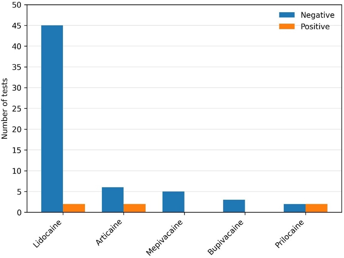
Overall, 56 children were tested with a single LA, 4 children with two different LAs, and 1 child with three LAs. Across the 61 included patients, a total of 67 skin prick tests were performed using full-strength LAs. All tested agents were amide-type LAs. The distribution of tested agents was as follows: lidocaine (n=47; 70.1%), articaine (n=8; 11.9%), mepivacaine (n=5; 7.5%), prilocaine (n=4; 6.0%), and bupivacaine (n=3; 4.5%).
LA allergy was confirmed in 5 of 61 children (8.2%) based on positive skin testing, with positivity observed on skin prick testing in 4 patients and on intradermal testing in 2 patients. Two patients showed hypersensitivity to articaine (P4, P5), one patient to prilocaine (P1), one patient to lidocaine (P3), and one patient to both prilocaine and lidocaine (P2) (Table 2). In all patients with confirmed LA allergy, safe alternative agents were identified using negative results from both skin testing (SPT/IDT) and subcutaneous DPT. SPTs were negative in 57 patients. Among these, two patients had positive IDT results at the 1:100 dilution and therefore did not proceed to provocation. Based on these findings, the negative predictive value of the 1:1 SPT was calculated as 96.4%. In patients with negative SPT and IDT results, no positivity was detected in subcutaneous provocation tests.
| P= Patient, M= Male, F= Female, LA= Local anesthetic, SPT= Skin prick test, IDT= Intradermal test, DPT= Drug provocation test, N/A= Not applicable. | |||||||
| Table 2. Clinical characteristics and test results of the patients with allergic reactions to LA. | |||||||
| Patient No |
|
Referral Clinical Finding |
|
|
|
|
|
| P1 |
|
Allergic reaction to non-LA drug |
|
|
|
|
|
|
|
|
|
|
||||
| P2 |
|
Anaphylaxis with LA (Lidocaine) |
|
|
|
|
|
|
|
|
|
|
||||
|
|
|
|
|
||||
| P3 |
|
Anaphylaxis with LA (Lidocaine) |
|
|
|
|
|
|
|
|
|
|
||||
| P4 |
|
Anaphylaxis with LA (Articaine) |
|
|
|
|
|
|
|
|
|
|
||||
| P5 |
|
Urticaria with LA (Unknown LA) |
|
|
|
|
|
|
|
|
|
|
||||
An exploratory sensitivity analysis was also performed by restricting the cohort to children with a genuine index reaction compatible with immediate LA hypersensitivity (n=18). In this subgroup, confirmed LA allergy was detected in 4 patients. Among children without an LA reaction history, 1 of 43 patients (2.3%) had confirmed LA allergy; this patient had been referred because of hypersensitivity to a non-LA drug. No confirmed LA allergy was detected among children referred solely for asthma, food allergy, or chronic urticaria (0/27).
Comparative analysis and risk factors
Children with confirmed LA allergy (n=5) were compared with those in whom LA allergy was not confirmed (n=56). Age at referral and sex distribution were comparable between groups, and no meaningful differences were observed with respect to accompanying allergic diseases or a family history of atopy.
Four of the children with true LA allergy were referred with a history of immediate hypersensitivity to LA administration. The rate of confirmed LA allergy was significantly higher in patients presenting with a history of suspected local anesthetic allergy compared with other referral reasons (p=0.024; OR 12, 95% CI 1.2–116) (Table 3). A suspected history of anaphylaxis with LA was more frequently recorded among children with true LA allergy and was associated with true LA allergy in the comparative analysis (p=0.005; OR 26.5, 95% CI 3.1–223.9) (Table 3).
| AEC, absolute eosinophil count; IQR, interquartile range; IgE, immunoglobulin E; LA, local anesthetic; OR, odds ratio. | |||
| Table 3. Comparison of clinical characteristics between children with and without confirmed LA allergy. | |||
| Data |
|
|
|
| Age (year), median (IQR) |
|
|
|
| Sex, n (%) |
|
||
| Female |
|
|
|
| Male |
|
|
|
| Parental allergic disease, n (%) |
|
|
|
| Atopy history of the children, n (%) |
|
|
|
| Concomitant allergic disease, n (%) | |||
| Asthma |
|
|
|
| Drug allergy |
|
|
|
| Food allergy |
|
|
|
| Urticaria |
|
|
|
| Atopic dermatitis |
|
|
|
| Allergic rhinitis |
|
|
|
| Referral reason, n (%) |
|
||
| Allergy to LA |
|
|
|
| Allergy to non-LA drug |
|
|
|
| Asthma |
|
|
|
| Food allergy |
|
|
|
| Chronic urticaria |
|
|
|
| Previous allergic reaction suspect to LA, n (%) |
|
|
|
| Previous Anaphylaxis suspect to LA, n (%) |
|
|
|
| AEC (cells/µL), median (IQR) |
|
|
|
| Eosinophil (%), median (IQR) |
|
|
|
| Total IgE (IU/mL), median (IQR) |
|
|
|
Of the 16 patients referred because of suspected hypersensitivity to non-LA drugs, one was diagnosed with confirmed LA allergy. Concomitant non-LA drug allergy was present in 2 of 5 (40.0%) children with confirmed LA allergy and in 12 of 56 (21.4%) children without confirmed LA allergy; however, this difference was not statistically significant (p=0.322). No test positivity or true allergy was detected in patients referred for other allergic conditions such as asthma, food allergy, or urticaria. Atopy did not differ significantly between the groups (p=0.127). When the drugs tested were examined, prilocaine or articaine was also associated with true LA allergy (p=0.012) (Figure 1).
DISCUSSION
In this 10-year retrospective study of children referred from dental clinics, confirmed immediate-type hypersensitivity to amide LAs was identified in a small percentage (8.2%) of fully evaluated children. Most referrals were prompted by co-existing allergic conditions rather than a history of LA-related reactions. True LA allergy was confirmed in only 5 of 61 fully evaluated children, and a safe alternative LA was successfully identified in every case using a stepwise diagnostic approach combining skin testing and subcutaneous DPT. Four of the five confirmed cases had a prior history of suspected LA allergy, while one had been referred because of hypersensitivity to a non-LA drug. Notably, true LA allergy was almost never detected in children without a history of an LA-related reaction, identifying this group as low-risk. Comparative analysis showed that a history of anaphylaxis and testing involving articaine or prilocaine were significantly associated with confirmed allergy, although these findings should be interpreted with caution given the small number of positive cases.
Pediatric literature over the past decade consistently demonstrates that true immediate-type LA allergy is rare among referred children.5-8 Recent real-world series report confirmation rates ranging from 1.3% to 4.3%, with most index events attributed to non-allergic mechanisms such as anxiety, vasovagal reactions, or dental pathology rather than the anesthetic itself.5-8 In the largest pediatric cohort from our country (n=231), Ocak et al. confirmed true LA allergy in 4.3% of cases, with mucocutaneous symptoms predominating.8 Similar diagnostic yields were reported by Aslan et al., Caliskan et al., and Selmanoglu et al.5-7 International data further support this observation; Kvisselgaard et al. found no IgE-mediated cases among 162 Danish patients, while Hascoët et al. estimated the incidence of anaphylaxis to LAs in dental practice at <0.01%.11,12 Our confirmation rate of 8.2% is slightly higher than most pediatric series. This difference likely reflects our selected population: we included only children with completed diagnostic work-up and adequate documentation, and all referrals originated from a tertiary dental service managing more complex cases.5-8
The low rate of confirmed LA allergy in the literature is largely explained by frequent misattribution of symptoms to the LA when other causes—such as non-LA drugs, disinfectants, latex, or procedural factors—are responsible.13,14 Studies evaluating LA allergy have reported that non-LA causes constitute 1.7-32% of all suspected reactions, depending on the procedure.15-17 Vasovagal reactions, hyperventilation, anxiety attacks, or toxicity symptoms can mimic allergy, but skin symptoms are usually absent.16,17 Despite the low rate of confirmed allergy, children with a suggestive history of immediate reaction to LA warrant thorough evaluation, as recommended by EAACI/ENDA guidelines, particularly when future exposure is anticipated.18
Clinical history alone cannot reliably differentiate true hypersensitivity from non-allergic reactions; therefore, objective testing is essential. SPT followed by IDT and DPT remain the cornerstone of diagnosis, whereas specific IgE assays and basophil activation tests are not widely used for LA reactions.17-19 Current literature recommends performing skin tests with preservative-free solutions and without vasoconstrictors such as epinephrine.15 Various IDT dilution protocols have been described.5-8,20 In our practice, IDT was performed at a 1:100 dilution using preservative- and vasoconstrictor-free preparations to minimize irritant false-positive results, and children with positive IDT were not challenged with the same agent.5-8,20 This cautious approach, together with the tertiary nature of our referrals, may partly explain our slightly higher confirmation rate compared with studies using 1:10 IDT dilutions.6,9,18-21 Subcutaneous DPT, performed in two graded steps at full strength, enabled identification of safe alternatives in all confirmed cases.22-27
The negative predictive value of undiluted SPT in our high-risk pediatric cohort was 96.4%, and no positive DPT results were observed after negative skin tests, consistent with previous reports.6,22,24,28 An important practical outcome of our study was the successful identification of a tolerated alternative LA in every child with confirmed allergy. This finding aligns with previous reports demonstrating that cross-reactivity among amide LAs is neither universal nor predictable.20,29 Although articaine and prilocaine showed higher positivity rates in our cohort, individualized testing allowed safe alternatives (most frequently mepivacaine) to be found in all cases, in line with EAACI/ENDA recommendations.18
Consistent with recent pediatric evidence, our findings indicate that the strongest predictors of true LA allergy are a convincing history of immediate reaction—particularly anaphylaxis—rather than background atopy or unrelated allergic comorbidities.6,8,11,30,31 Age, sex, family history of atopy, and co-existing allergic diseases did not differ between allergic and non-allergic groups. In contrast, history of anaphylaxis to LA and testing with articaine or prilocaine emerged as key risk signals. These observations parallel the findings of Ocak et al.8 The role of hypersensitivity to other drugs is less clear. Some adult data suggest that multiple drug allergies or concomitant drug allergies may be more common in patients with positive LA test results.20 Suleyman et al. found no significant difference between patients with LA allergy and the control group in terms of confirmed drug allergy to NSAIDs or β-lactams.30 A study by Kemoklidze et al., involving 450 pediatric and adult patients, found that drug allergy co-occurring with food allergy was associated with an increased risk of LA allergy.32 Importantly, suspected hypersensitivity to a non-LA drug as a referral indication should be distinguished from concomitant non-LA drug allergy as a comorbidity. In our cohort, concomitant drug allergy was numerically more frequent among children with confirmed LA allergy, although this difference was not statistically significant. Previous studies have suggested a possible association between concomitant drug allergies and LA hypersensitivity; however, the available evidence remains inconsistent, and larger studies are needed to clarify whether concomitant drug allergy represents an independent risk factor. Importantly, our data should not be interpreted as supporting routine pre-procedural LA skin testing in children with atopy or allergic comorbidities alone. In our cohort, these low-risk referrals reflected institutional and referring-physician practice during the study period. The absence of confirmed LA allergy in this subgroup supports counseling dentists and families that unrelated allergic comorbidities alone are not an indication for routine LA testing. In summary, this study confirms that true LA allergy is infrequent but not negligible in children referred from dentistry. A history of immediate reaction, especially anaphylaxis, identifies a high-yield subgroup that benefits from formal allergologic evaluation, whereas routine testing appears unnecessary in low-risk children referred for other allergic conditions. These results support the implementation of risk-stratified diagnostic algorithms to optimize resource use while ensuring patient safety.
This study has several limitations. Its retrospective, single-center design and tertiary dental referral setting may limit generalizability and introduce selection bias. The number of confirmed cases was small, resulting in wide confidence intervals and precluding multivariable analysis. Some records lacked detailed information on index symptoms or subsequent dental exposures, and the diagnostic approach evolved within institutional practice over the 10-year period. In addition, some children without a previous LA reaction underwent IDT because of pre-procedural referral requests; although testing was performed with diluted, vasoconstrictor-free preparations under direct supervision, the theoretical risk of iatrogenic sensitization cannot be completely excluded. These limitations further support cautious interpretation and a risk-stratified approach.
In conclusion, although immediate-type allergy to local anesthetics is rare in children, it can occur and may involve cross-reactivity between agents. Structured diagnostic testing combining skin tests and drug provocation is essential for accurate confirmation or exclusion of hypersensitivity and for identifying safe alternatives. Many evaluations, however, are performed unnecessarily in low-risk patients. A risk-based approach—prioritizing children with a history of immediate reaction to LA, particularly anaphylaxis —should guide clinical decision-making. The development of standardized, risk-stratified diagnostic pathways has the potential to reduce unnecessary testing, decrease parental anxiety, and facilitate timely dental care without compromising safety.
Ethical approval
This study was approved by the Dr. Behçet Uz Pediatric Diseases and Surgery Training and Research Hospital Institutional Local Ethics Committee (Date: 12 Mar 2026, Decision/Protocol No: GOA-319). Informed consent was obtained from all participants involved in this study.
Data availability statement
The data that support the findings of this study are available from the corresponding author upon reasonable request.
Conflict of interest
The authors declare that this study was conducted in the absence of any commercial or financial relationships that could be construed as a potential conflict of interest.
Funding
The authors declare that this study received no funding.
Generative AI statement
The authors declare that during the preparation of this study, the following AI-assisted technology was used: ChatGPT (OpenAI) on Not specified. Extent of Use: English language editing, including improvements in grammar and fluency. The authors confirm that they have critically reviewed and edited any AI-generated content and take full responsibility for the integrity, accuracy, and originality of the publication. The authors certify that the original human contribution is maintained and that AI-assisted tools are not listed or cited as authors.
References
- American Academy of Pediatric Dentistry. Use of local anesthesia for pediatric dental patients. In: The Reference Manual of Pediatric Dentistry. Chicago (IL): American Academy of Pediatric Dentistry; 2024: 386-93.
- Wieshuber C, Stoevesandt J, Trautmann A. The needle in the haystack: allergic anaphylaxis caused by the local anesthetic articaine. Eur Ann Allergy Clin Immunol. 2014;46:38-40.
- Gall H, Kaufmann R, Kalveram CM. Adverse reactions to local anesthetics: analysis of 197 cases. J Allergy Clin Immunol. 1996;97:933-7. https://doi.org/10.1016/s0091-6749(96)80067-4
- Cuesta-Herranz J, de las Heras M, Fernández M, et al. Allergic reaction caused by local anesthetic agents belonging to the amide group. J Allergy Clin Immunol. 1997;99:427-8. https://doi.org/10.1016/s0091-6749(97)70064-2
- Caliskan N, Yildirim G, Bologur H, et al. Local anesthetics allergy in children: Evaluation of diagnostic tests with Real-Life data. Pediatr Allergy Immunol. 2024;35:e14097. https://doi.org/10.1111/pai.14097
- Aslan S, Anıl H, Kaya M, Harmancı K. Evaluation of diagnostic tests for immediate-type allergic reactions to amide group local anesthetics in children. Pediatr Allergy Immunol. 2025;36:e70085. https://doi.org/10.1111/pai.70085
- Selmanoglu A, Güvenir H, Celik IK, et al. Immediate local anesthetic reactions and diagnostic test results in pediatric patients. Allergol Immunopathol (Madr). 2021;49:108-14. https://doi.org/10.15586/aei.v49i3.87
- Ocak M, Bal Çetinkaya F, Akarsu A, et al. Thirty years’ experience with immediate-type local anesthetic hypersensitivity reactions in children. Pediatr Allergy Immunol. 2025;36:e70178. https://doi.org/10.1111/pai.70178
- Cardona V, Ansotegui IJ, Ebisawa M, et al. World allergy organization anaphylaxis guidance 2020. World Allergy Organ J. 2020;13:100472. https://doi.org/10.1016/j.waojou.2020.100472
- Brockow K, Garvey LH, Aberer W, et al. Skin test concentrations for systemically administered drugs - an ENDA/EAACI Drug Allergy Interest Group position paper. Allergy. 2013;68:702-12. https://doi.org/10.1111/all.12142
- Kvisselgaard AD, Mosbech HF, Fransson S, Garvey LH. Risk of immediate-type allergy to local anesthetics is overestimated-results from 5 years of provocation testing in a Danish allergy clinic. J Allergy Clin Immunol Pract. 2018;6:1217-23. https://doi.org/10.1016/j.jaip.2017.08.010
- Hascoët E, Mahé J, Meillard H, Théophile H, Cloitre A, Lesclous P. Anaphylactic reactions to local anesthetics in dental practice: a nationwide French retrospective study. Clin Oral Investig. 2022;26:1667-76. https://doi.org/10.1007/s00784-021-04139-5
- Neal JM, Barrington MJ, Fettiplace MR, et al. The third American Society of Regional Anesthesia and Pain Medicine practice advisory on local anesthetic systemic toxicity: executive summary 2017. Reg Anesth Pain Med. 2018;43:113-23. https://doi.org/10.1097/AAP.0000000000000720
- Mertes PM, Malinovsky JM, Jouffroy L, at al. Reducing the risk of anaphylaxis during anesthesia: 2011 updated guidelines for clinical practice. J Investig Allergol Clin Immunol. 2011;21:442-53.
- Harboe T, Guttormsen AB, Aarebrot S, Dybendal T, Irgens A, Florvaag E. Suspected allergy to local anaesthetics: follow-up in 135 cases. Acta Anaesthesiol Scand. 2010;54:536-42. https://doi.org/10.1111/j.1399-6576.2009.02193.x
- Kvisselgaard AD, Krøigaard M, Mosbech HF, Garvey LH. No cases of perioperative allergy to local anaesthetics in the Danish Anaesthesia Allergy Centre. Acta Anaesthesiol Scand. 2017;61:149-55. https://doi.org/10.1111/aas.12833
- Trautmann A, Goebeler M, Stoevesandt J. Twenty years’ experience with anaphylaxis-like reactions to local anesthetics: genuine allergy is rare. J Allergy Clin Immunol Pract. 2018;6:2051-58.e1. https://doi.org/10.1016/j.jaip.2018.04.005
- Barbaud A, Garvey LH, Torres M, et al. EAACI/ENDA position paper on drug provocation testing. Allergy. 2024;79:565-79. https://doi.org/10.1111/all.15996
- Malinovsky JM, Chiriac AM, Tacquard C, Mertes PM, Demoly P. Allergy to local anesthetics: reality or myth? Presse Med. 2016;45:753-7. https://doi.org/10.1016/j.lpm.2016.05.011
- YIlmaz I, Özdemir SK, Aydin Ö, Çelik GE. Local anesthetics allergy: who should be tested? Eur Ann Allergy Clin Immunol. 2018;50:66-71. https://doi.org/10.23822/EurAnnACI.1764-1489.38
- Demoly P, Adkinson NF, Brockow K, et al. International consensus on drug allergy. Allergy. 2014;69:420-37. https://doi.org/10.1111/all.12350
- Specjalski K, Kita-Milczarska K, Jassem E. The negative predictive value of typing safe local anesthetics. Int Arch Allergy Immunol. 2013;162:86-8. https://doi.org/10.1159/000350763
- Jiang S, Tang M. Allergy to local anesthetics is a rarity: review of diagnostics and strategies for clinical management. Clin Rev Allergy Immunol. 2023;64:193-205. https://doi.org/10.1007/s12016-022-08937-x
- McClimon B, Rank M, Li J. The predictive value of skin testing in the diagnosis of local anesthetic allergy. Allergy Asthma Proc. 2011;32:95-8. https://doi.org/10.2500/aap.2011.32.3417
- Astarita C, Gargano D, Romano C, et al. Long-term absence of sensitization to mepivacaine as assessed by a diagnostic protocol including patch testing. Clin Exp Allergy. 2001;31:1762-70. https://doi.org/10.1046/j.1365-2222.2001.01226.x
- Chandler MJ, Grammer LC, Patterson R. Provocative challenge with local anesthetics in patients with a prior history of reaction. J Allergy Clin Immunol. 1987;79:883-6. https://doi.org/10.1016/0091-6749(87)90236-3
- Tomoyasu Y, Mukae K, Suda M, et al. Allergic reactions to local anesthetics in dental patients: analysis of intracutaneous and challenge tests. Open Dent J. 2011;5:146-9. https://doi.org/10.2174/1874210601105010146
- Koca Kalkan I, Koycu Buhari G, Ates H, et al. Identification of risk factors and cross-reactivity of local anesthetics hypersensitivity: analysis of 14-years’ experience. J Asthma Allergy. 2021;14:47-58. https://doi.org/10.2147/JAA.S292442
- Halling F, Neff A, Meisgeier A. True allergies to articaine: a 25-year analysis. Dent J (Basel). 2025;13:180. https://doi.org/10.3390/dj13050180
- Suleyman A, Tamay Z, Guler N. Risk factors for immediate-type local anesthetic hypersensitivity reactions in pediatric patients: a retrospective case-control study. J Asthma Allergy. 2022;15:453-60. https://doi.org/10.2147/JAA.S349637
- Cetinkaya F. Sensitivity to local anaesthetics among asthmatic children. Int J Paediatr Dent. 2001;11:405-8.
- Kemoklidze T, Topuridze N, Mchedlishvili L, Kamkamidze G. Risk factors influencing hypersensitivity to the local anesthetic drugs. Georgian Med News. 2022;5:62-5.
Copyright and license
Copyright © 2026 The author(s). This is an open-access article published by Aydın Pediatric Society under the terms of the Creative Commons Attribution License (CC BY) which permits unrestricted use, distribution, and reproduction in any medium or format, provided the original work is properly cited.